5-in-1 System: 3D OCT, Fundus Camera, Angiography, Biometry, Topography
- 5-in-1 System : 3D OCT, Fundus Camera, Angiography, Biometry, Topography
- 80,000 A-scan/sec Quick Capture, Clear Answer
- True Color Fundus in One Shot
- Enhanced Angiography Fast OCT-A Imaging, Full Coverage
- Precise and Layered Analysis with Biometry & Topography
- Accurate Analysis
Features
5 in 1 System : 3D OCT, Fundus Camera, Angiography, Biometry, Topography
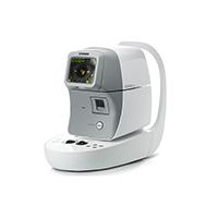
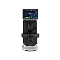
OCTavius integrates five essential diagnostic functions—3D OCT, Fundus Camera, Angiography, Biometry, and Topography—into a single device. It offers seamless operation and delivers precise results needed for clinical decision-making. With the 5-in-1 system, enhance your diagnostic accuracy while maximizing workflow efficiency and exam room space.
80,000 A-scan/sec Quick Capture, Clear Answer
Even with involuntary eye movements or blinking, OCTavius captures stabilized, high-resolution 3D images through ultra-fast scanning at up to 80,000 A-scans per second. This ensures clear, repeat-free imaging—ideal even for beginners. Visualized retinal layers allow clinicians to observe pathological structures such as layer thickness and macular differentiation with greater clarity and confidence.
True Color Fundus in One Shot
With 12-bit color depth and gamma correction, OCTavius captures true color fundus images in a single shot—free from color distortion. It accurately balances contrast across both dark retinal areas and bright optic discs, delivering clear visualization of arteries, veins, and even fine microvasculature. Additionally, it automatically aligns and merges multiple fundus images—up to seven—to generate a widefield panoramic view, enabling intuitive identification of lesion location and extent in a single, comprehensive image.
Enhanced Angiography Fast OCT-A Imaging, Full Coverage
OCTavius offers multiple scan sizes—3×3, 4.5×4.5, 6×6, and 9×9mm²—allowing clinicians to tailor vascular imaging based on lesion location and extent. From localized detail to widefield coverage, it enables efficient and precise visualization of microvasculature in a single, dye-free scan. This reduces diagnostic burden while enhancing workflow efficiency.
Detailed measurement of biometry and topography
OCT Topography enables simultaneous measurement of both anterior and posterior corneal surfaces, delivering detailed, three-dimensional analysis. With 16 types of corneal maps, clinicians can assess corneal thickness, curvature, and elevation changes with precision. Optical Biometry visualizes the full axial structure—from cornea to macula—in high-resolution 2D, providing more detailed data than traditional ultrasound. This supports accurate IOL selection tailored to each patient’s ocular profile.
Accurate Analysis
Through integrated analysis, it's possible to grasp personalized symptoms, diseases, and their progression for each patient at a glance. This includes tracking pathological changes with Progression analysis, comparing pre- and post-treatment states with Compare functionality, analyzing pathological conditions from various perspectives such as key indicators compared to normative data.
Brochures, Guides, and Documents
Get in touch with us now!
1800 251 852
info@opticare.com.au
NSW 2144
Phone: 02 9748 8777
Fax: 02 9748 8666
QLD 4011
Phone: 07 3630 2366
Fax: 07 3630 2399
Wangara, WA 6065
Phone: 08 9376 3700
Check our other diagnostic solutions
Interested to know more about the Huvitz OCTavius all-in-one OCT?
Your questions are all welcome. Contact us and we'll be in touch right away.